
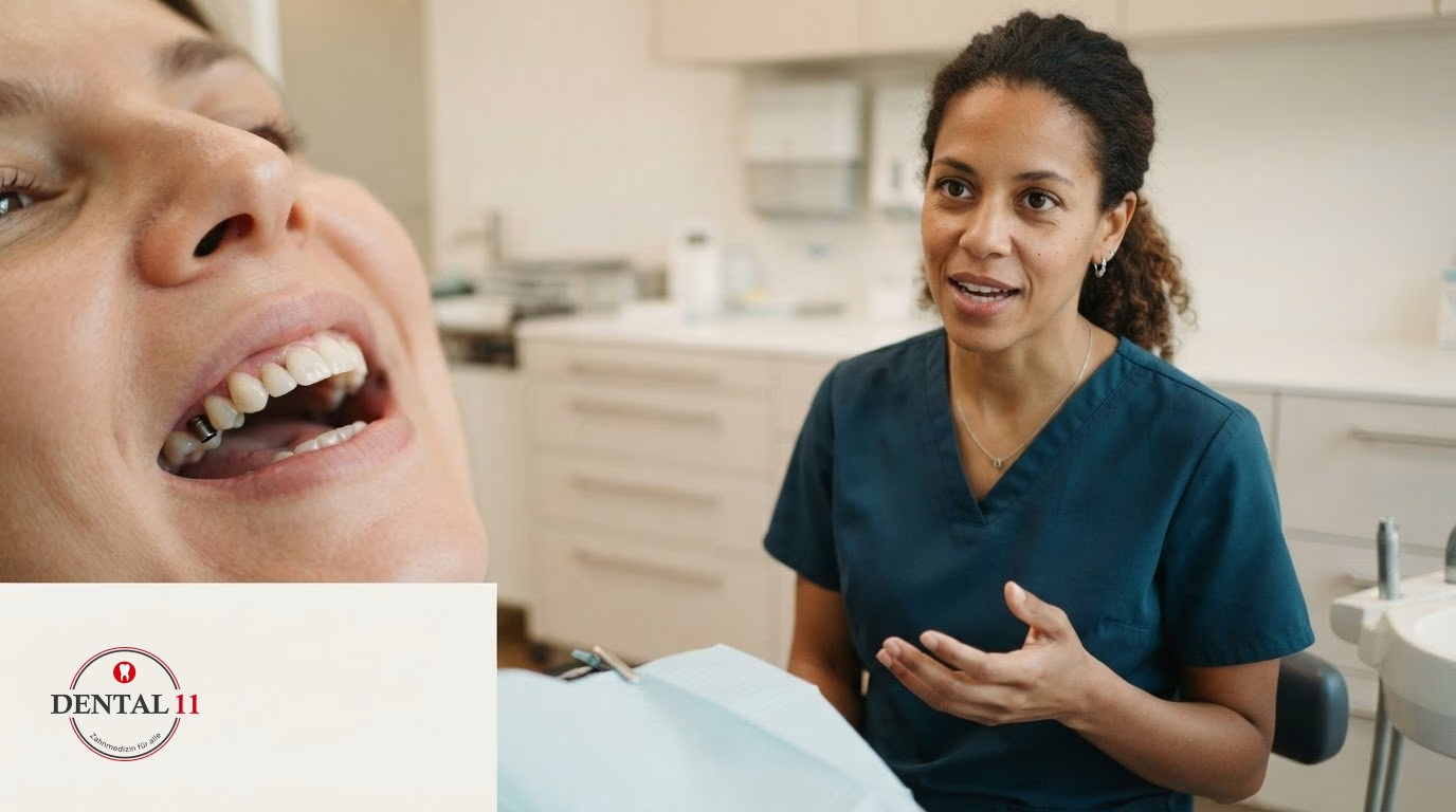
More gum tissue over an implant does not automatically mean that something is wrong. If the gum grows over an implant, this can be part of the healing process. However, persistent redness, tenderness or bleeding should be assessed by a dentist.
A red area can be unsettling, especially shortly after the implant has been placed. What matters is whether the symptoms ease or get worse and whether other changes appear. Peri-implantitis cannot be identified from the colour of the gum alone.
Here you will learn which signs may point to inflammation around an implant and how peri-implantitis differs from periodontitis. You will also find out why healthy, well cared for gums are important for keeping the implant in the long term and what a dental assessment involves. This helps you put changes into context without diagnosing redness yourself too quickly.
Key points
- A change at the implant is easier to put into context if you pay attention to how it develops and to additional signs such as bleeding or tenderness.
- Healthy gums and careful cleaning help protect the tissue around the implant.
- Excess tissue can have different causes. Which treatment makes sense depends on the findings at the implant and the gum.
- Peri-implantitis affects the tissue around the implant, periodontitis the supporting structures of natural teeth. A dental examination helps to tell these changes apart.
Why healthy gums matter for implants
Healthy gums protect the tissue around an implant and make daily cleaning easier. In the first days after the procedure, a blood clot forms that contributes to healing. Gentle brushing protects this clot and the sensitive area.
Swelling and mild signs of inflammation can occur in the early healing phase. Swelling may go down about a week after the implant is placed, but healing varies from person to person. Even if the gum still feels soft, this can be part of the healing process. Slight bleeding when brushing is possible during healing. What matters is whether the symptoms improve or get worse. Persistent or increasing symptoms should be assessed by a dentist.
The soft tissue is also important for the next steps of the restoration. The crown can be fitted once the gum is stable and firmly attached. At the same time, osseointegration begins: bone tissue bonds with the surface of the implant. Gum and bone have different roles, but both need to be monitored carefully.
A change in the shape of the gum is not automatically a sign of disease. If gum grows over the implant, this may be related to healing or to how the tissue develops. An examination shows whether the tissue is free of irritation and easy to clean. Pain alone does not reliably show whether the tissue around the implant is healthy.
Regular toothbrushing, cleaning between the teeth and professional check-ups are important for caring for the area around implants. Attention is also paid to plaque in places that are hard to reach. This way, changes to the gum or to the implant restoration can be spotted early, before they make cleaning more difficult.
When should you see a dentist about gum problems at an implant? If there is inflammation, persistent redness, bleeding, swelling or pain, a dental assessment should take place. An examination can clarify whether only the gum is affected or whether deeper tissue also needs to be checked.
Stable, firmly attached soft tissue is important for securely anchoring the crown. The gum around dental implants still needs regular care and cleaning.
Redness and tenderness of the gum
Colour alone does not reliably show what is causing a change at the implant. Findings on touch and the dental examination are also important for putting it into context.
Redness and tenderness to pressure
Redness and tenderness to pressure are changes in the gum tissue that you can see or feel. They can point to local irritation or an inflammatory reaction, but on their own they do not prove peri-implantitis. For this reason, do not judge a change by its colour alone. The exact location and how it develops also give the dentist important clues.
During the examination, the dentist may carry out what is known as a BOP test (bleeding on probing). The gum is gently probed with a periodontal probe to see whether it starts to bleed. The test adds to the visual findings but does not replace further assessment. A single observation does not yet reliably show how deeply the tissue is affected.
At your appointment, describe as precisely as possible where you feel the tenderness and whether it changes. This information helps to examine the affected area in a targeted way. If available, earlier findings can also be useful for comparison.
Inflammation and gum recession
The tissue around the implant can become inflamed or recede. A change in the gum margin can affect the shape of the transition between the tissue and the restoration. To put this into context, it is important to know whether only the visible mucosa looks unusual or whether there are further findings.
For this reason, the examination looks not only at redness but also at the contour of the tissue and the area around the implant. A tender area can have different causes; neither the cause nor the extent can be deduced from how it feels alone. The dental assessment combines your description with the clinical observations so that the change can be put into context in a targeted way.
How is excess gum tissue at an implant treated?
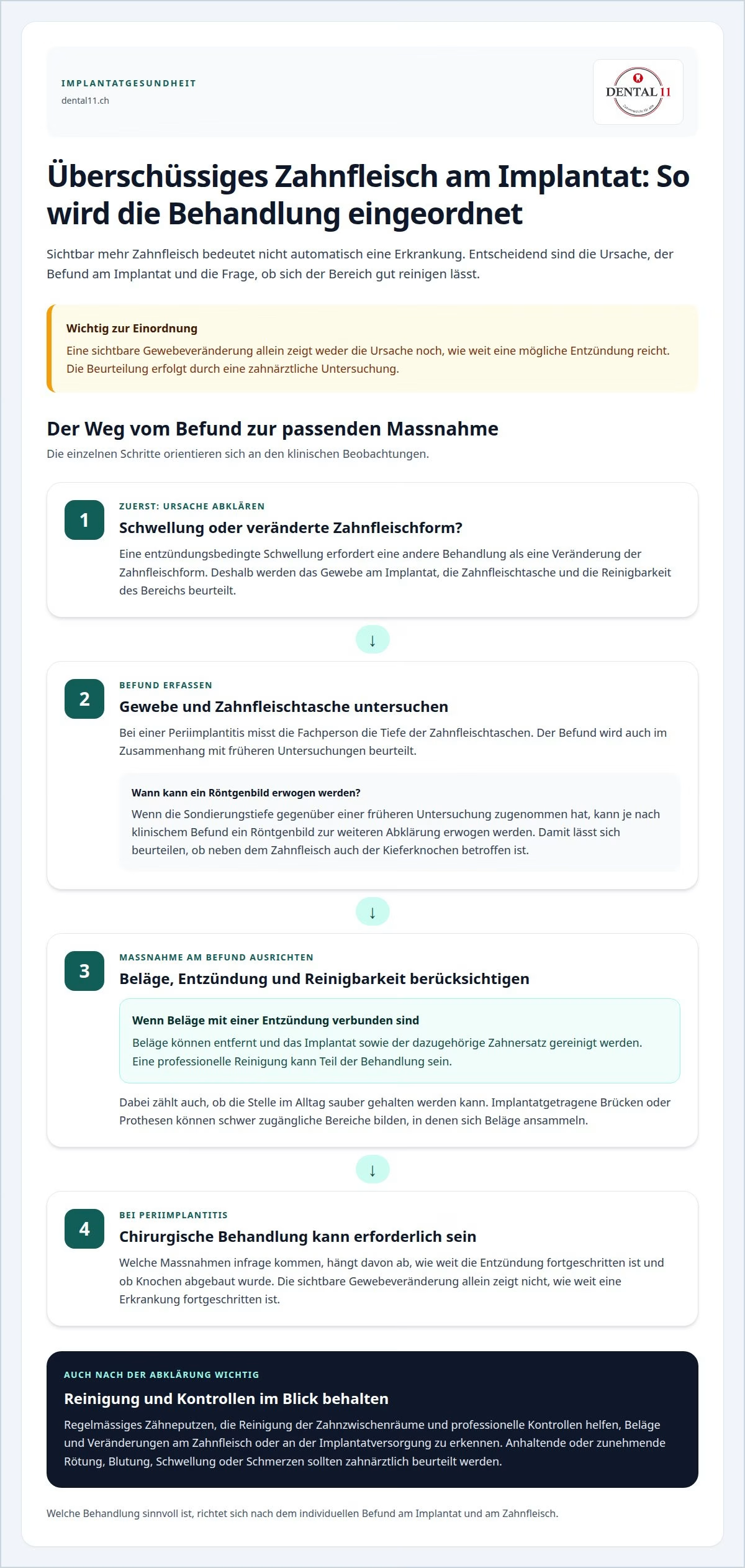
Before any treatment, the dentist clarifies why the tissue appears enlarged. Swelling caused by inflammation needs a different treatment from a change in the shape of the gum. The tissue around the implant, the gum pocket and how easily the area can be cleaned are therefore examined.
In the case of peri-implantitis, the dentist measures the depth of the gum pockets. If the probing depth has increased compared with an earlier examination, an X-ray may be considered for further assessment, depending on the clinical findings. This helps to assess whether the jawbone is affected as well as the gum. A visible change in the tissue alone does not show how far the disease has progressed.
If plaque is linked to inflammation, it can be removed and the implant and its restoration cleaned. Professional cleaning can be part of the treatment. It also matters whether the affected area can be kept clean in everyday life. Implant-supported bridges or dentures can create areas that are hard to reach, where plaque builds up.
Peri-implantitis may require surgical treatment. Which measures are suitable depends on how far the inflammation has progressed and whether bone has been lost. If there is a pronounced bone defect, oral surgical bone augmentation may be necessary. Bone substitute material or the patient’s own bone can be used for this.
Gum grafting is a different treatment: it targets receded tissue, not excess tissue. If there is enough bone, the soft tissue can be thickened with the patient’s own tissue or a tissue matrix. The patient’s own tissue is often taken from the palate; a dermal tissue matrix can avoid this removal. Which method is suitable depends on the findings and the aim of treatment.
Do not remove the tissue yourself, as this can injure the implant and the surrounding tissue. Sharp instruments or harsh products can also irritate the area. A professional assessment clarifies whether cleaning, treatment of the inflammation or a surgical procedure is needed.
Can the gum around an implant become inflamed or recede?
The tissue around an implant can change because the transition between the mucosa and the implant is structured differently from that between the gum and a natural tooth. The seal formed there is not as tight. This is why caring for the transition area is particularly important, especially when the restoration makes access more difficult.
For the dental assessment, the visible outline of the tissue is not the only factor. The dentist probes the gum pocket and compares the measurement with earlier check-ups. An increased probing depth of around 5 to 6 millimetres can be a reason for further assessment. If the depth has increased since the last examination, an X-ray can help to assess the bone area.
The shape of the restoration also influences daily care. Spaces under implant-supported bridges or dentures can be hard to reach. If the design leaves little access for cleaning, plaque can be more difficult to remove there. At check-ups, the dentist can assess whether the restoration is easy to clean and which areas need particular attention when caring for it.
A professional assessment takes into account measurements, earlier findings and the condition of the restoration. It helps to distinguish whether the mucosa has changed or whether a more in-depth assessment is needed. If you have questions or notice changes, you can arrange a dental assessment. This gives the examination a concrete starting point and allows it to be tailored to your implant situation.
The gum can grow over dental implants or recede. Inflammation of the mucosa around an implant is first called peri-implant mucositis and can develop into peri-implantitis. Inflammation in the area of the implant can affect the gum or the tooth socket. The quality and mobility of the mucosa can influence cleaning and should be assessed together with other clinical factors. Poor oral hygiene can allow bacteria to settle in plaque and promote peri-implantitis.
If there is enough bone, receded gum around an implant can be treated by thickening the soft tissue with the patient’s own tissue or a tissue matrix. Various aspects of the tissue situation are taken into account: growth or recession of the gum, the quality of the mucosa and any inflammation. The terms distinguish inflammation of the mucosa from inflammation of the gum or tooth socket. Whether receded tissue can be treated depends on the amount of bone available; the options are the patient’s own tissue or a tissue matrix.
Assessment at Dental11 in Zurich Enge
Dental11 helps you have changes at your implant assessed professionally. If there is redness, bleeding or a change in the gum margin, an examination can clarify which tissues are affected and whether further assessment is needed. How easily the area around the implant and restoration can be cleaned can also be assessed.
Our care includes dental implants as well as prevention and professional teeth cleaning. Regular check-ups can help to spot changes at the implant early and adapt your care accordingly. Which treatment makes sense depends on the findings and your individual needs.
If you notice a change or have questions about caring for your implant, you can book an appointment for a dental assessment. Feel free to contact Dental11 so that your situation can be discussed calmly.
Get clarity about the next step
If the gum grows over an implant, make a note of when you first noticed the change and how it is developing. Also note whether bleeding, tenderness to pressure or other changes occur. These observations give the conversation at the dental practice a concrete starting point, but they do not replace an examination.
Your personal history and the current findings both count in the assessment. If available, bring information about earlier implant check-ups or changes to your restoration. This allows the dentist to put the course of events into context and discuss with you which further assessment or everyday care is suitable. The next sensible step depends on the clinical findings and your individual needs.
If you would like to discuss a change, contact Dental11. A calm, open conversation helps to answer your questions and plan the next step together.
Frequently asked questions about peri-implantitis
How can a patient recognise peri-implantitis?
A reliable diagnosis is not possible through self-observation. The dentist assesses the implant and the surrounding tissue; among other things, the gum pockets are probed to make a diagnosis. Comparing the results with earlier examinations can also help to put the findings into context. This makes it possible to distinguish changes at the implant from diseases of the supporting structures of natural teeth.
Can peri-implantitis be cured?
Peri-implantitis can be treated, but the outcome depends on how far the disease has progressed. The aim is to control inflammation and plaque and to stabilise the tissue around the implant. In advanced cases, surgical treatment may be needed. Dental follow-up care remains important in order to monitor the condition and adjust the treatment if necessary.
What happens as the gum heals after a dental implant is placed?
After an implant procedure, a blood clot forms first, which contributes to wound healing. The soft tissue then heals gradually while the bone bonds with the surface of the implant. How healing progresses depends on factors such as your individual starting situation, your general health and how you look after the area during healing. The treating practice accompanies this process with check-ups.
What are the symptoms of peri-implantitis?
Peri-implantitis can show itself in different ways and does not have to cause pain at first. The absence of pain is therefore not a reliable sign of healthy tissue. Persistent or recurring changes should be assessed by a dentist, even if they cause hardly any discomfort at first. Only an examination can clarify whether there is a peri-implant disease or another cause.
What causes peri-implantitis?
Peri-implantitis is linked to inflammation at the implant and can be promoted by bacterial plaque. Areas under bridges or dentures that are hard to reach can also make cleaning more difficult. Smoking, diabetes, osteoporosis and certain medications are considered possible risk factors. These factors do not automatically lead to disease in everyone, but they are relevant for the assessment.
How can peri-implantitis be prevented?
Careful oral care, cleaning between the teeth and regular dental check-ups are important. With implant-supported restorations, care should also include places that are hard to reach. The dentist can explain which areas of your restoration need particular attention. This way, daily cleaning can be tailored specifically to your implant, crown, bridge or denture.
This article is for general information and does not replace a personal dental examination.


